Assistant Professor · Computer Science & Engineering
IIIT Vadodara

Reading the genome
in the image.

I work at the intersection of data science, medical image analysis and oncology — building radiomic and radiogenomic models that link what a tumour looks like on a scan to what it is doing molecularly, so clinicians can characterise a cancer before a needle ever touches it.

Dept. of Computer Science & Engineering
Indian Institute of Information Technology Vadodara
Gandhinagar Campus, Gujarat, India
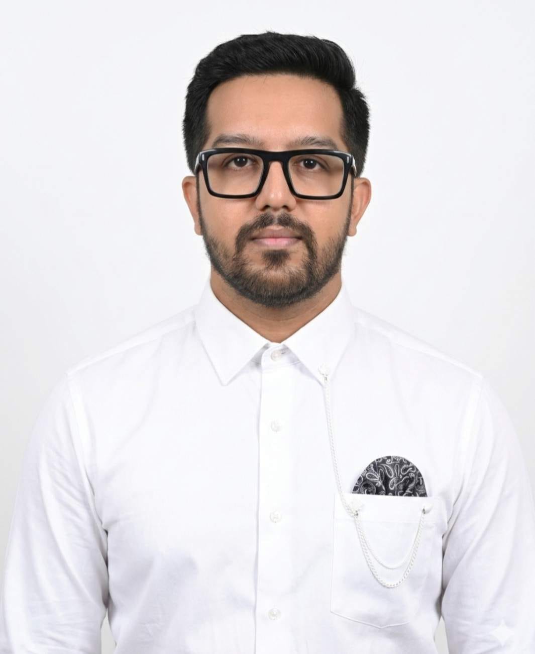
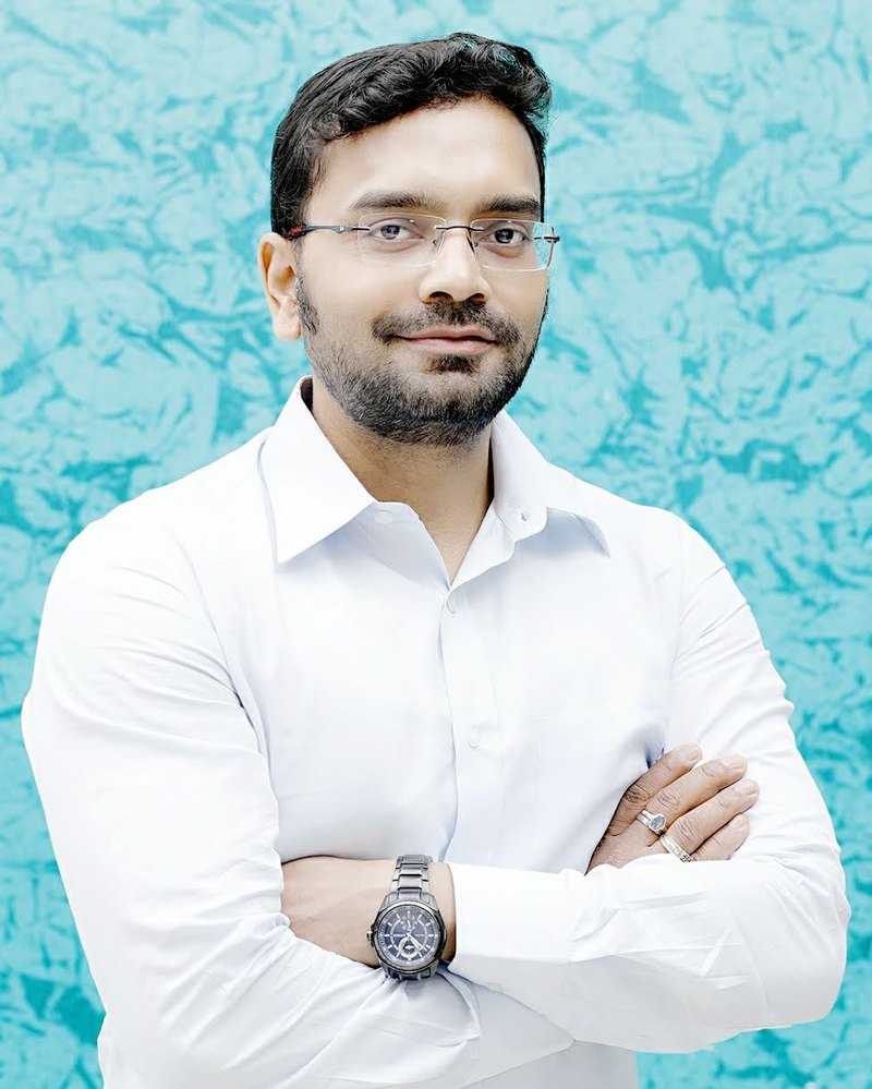
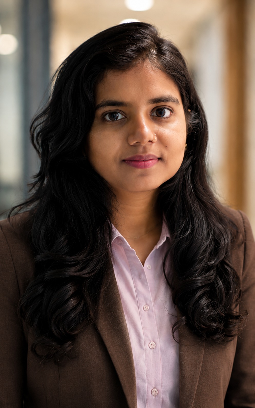
Portrait of Dr. Sanjay Saxena
Dr. Sanjay Saxena Ph.D. & M.Tech, IIT (BHU) Varanasi
Postdoctoral Research, University of Pennsylvania, USA
0h-index
0i10-index
0Citations
0Edited books
Top 2%Stanford/Elsevier 2025

Research

Four connected questions about
imaging, biology and scale.

Cancer care is moving from population averages to individual biology — but the molecular tests that guide treatment are invasive, expensive and vulnerable to sampling bias. My group asks whether imaging, which every patient already receives, can carry that same molecular signal. The answer increasingly is yes.

Radiomics & radiogenomics in oncology

Imaging as a molecular proxy

Quantifying tumour phenotype from routine MRI and CT, then mapping those signatures onto genotype — IDH mutation, MGMT promoter methylation, 1p/19q co-deletion — to make molecular characterisation non-invasive and repeatable across the treatment course.

Medical image analysis

Methods that hold up in the clinic

Segmentation, harmonisation across scanners and interpretable deep architectures for brain, liver, breast and vascular imaging. The emphasis is generalisation: a model that only works on the cohort it was trained on is not a clinical tool.

AI in healthcare

Federated, generative and clinically usable

Privacy-preserving federated learning so models can learn across hospitals without patient scans ever leaving the institution — plus large language models and digital twins for translational healthcare applications.

Data science

Scale, bias and imaging biomarkers

High-performance and parallel computing for large imaging cohorts, systematic bias evaluation of published AI pipelines, and the statistical work of turning a promising feature into a defensible imaging biomarker.

Recent research highlights

Selected work from 2025–26.

Neuroradiology2026 · Springer

The tumour margin carries the signal

Radiomic analysis usually stops at the visible tumour boundary. Working with multi-parametric MRI from over 500 glioblastoma patients, we extracted roughly 11,000 deep features per patient from tumour and progressively dilated peritumoral regions. Adding a 10 mm peritumoral margin lifted MGMT promoter methylation prediction from an AUC of 0.71 to 0.81, and an 8 mm margin improved survival prediction — evidence that the apparently normal tissue surrounding a glioblastoma encodes both prognostic and molecular information.

Read the paper →
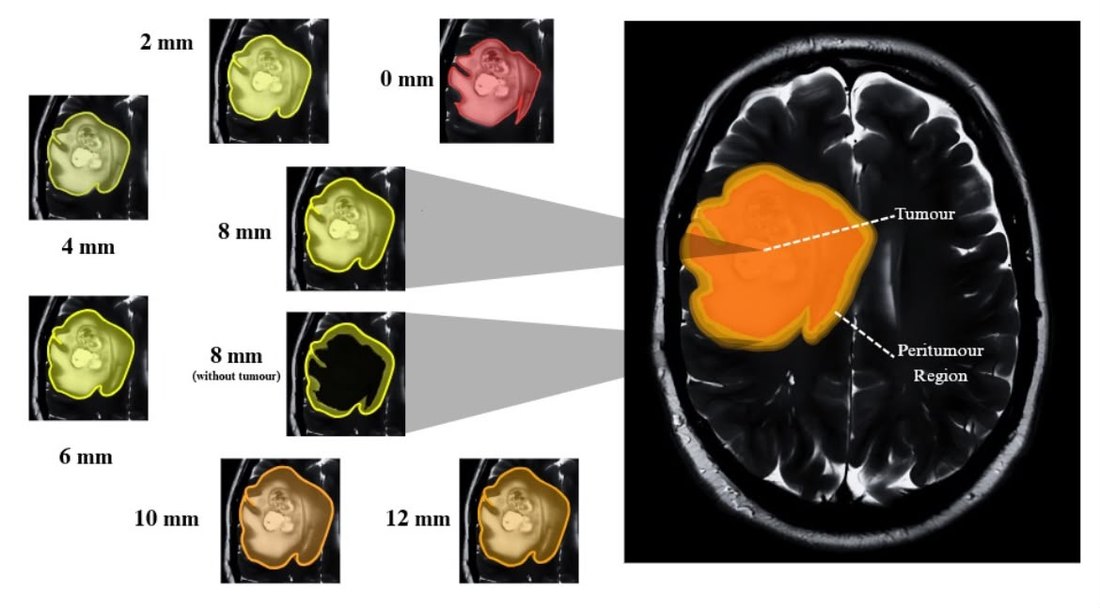
Progressively dilated peritumoral regions from 0 to 12 mm around a glioblastoma on T2 MRI, with tumour and peritumour regions annotated
Progressively dilated peritumoral margins (0–12 mm) analysed around the tumour core; the 8 mm ring is also shown with the tumour masked out.
Neuro-Oncology2025 · SNO Annual Meeting

Graphs and texture, working together

IDH-wildtype glioblastoma carries a median survival of 12–18 months, and existing survival models struggle with its spatial heterogeneity. We encoded segmented tumour subregions as graph structures using intra- and inter-slice connectivity, fused them with handcrafted texture descriptors and clinical variables, and validated across a 400-patient discovery cohort and a 128-patient replication cohort — a non-invasive route to survival estimation that respects how the tumour is actually arranged in space.

Read the abstract →
J. Medical & Biological Engineering2025 · Springer

RFiLM U-Net: letting radiomics steer the network

Rather than treating radiomic features as an output, we used them as a control signal. A first U-Net segments the liver from abdominal CT; radiomic descriptors extracted from that segmentation then linearly modulate a second network that refines tumour delineation. The two-stage design reached Dice coefficients of 0.92 for liver and 0.87 for tumour under five-fold cross-validation, outperforming the prior state of the art.

Read the paper →
Measurement2026 · Elsevier

Attention mechanisms for vascular risk

Extending the same methodological toolkit beyond oncology: attention- and transformer-based architectures for carotid artery segmentation and automated intima-media thickness and plaque area measurement in ultrasound scans — quantitative cardiovascular risk markers extracted from an imaging modality that is cheap, portable and already ubiquitous.

Read the paper →

Background

Varanasi to Philadelphia to Gandhinagar.

Clinical collaboration

Sustained joint work with AIIMS New Delhi, NIMHANS Bengaluru and Taipei Medical University, keeping method development anchored to real diagnostic questions and curated neuro-oncology cohorts.

Books & intellectual property

Five edited volumes with Elsevier, Taylor & Francis and IGI Global — including the two-volume Radiomics and Radiogenomics in Neuro-Oncology — and a German patent on a COVID-19 treatment recommender system.

Editorial & peer review

Guest Editor for Diagnostics. Reviewer for IEEE Transactions on Medical Imaging, Scientific Reports, Computers in Biology and Medicine, Biomedical Signal Processing & Control and Brain Informatics.

Teaching

  • Artificial Intelligence
  • Distributed & Parallel Computing
  • Data Analytics & Computing
  • Data Science for AI in Healthcare
  • Digital Image Processing
  • Data Structures
  • Scientific Computing with Python

Research students

The people doing the work.

Supervision spans quantum machine learning, federated healthcare systems and the full radiogenomics pipeline for glioblastoma. Several of the papers above began as a student thesis.

Doctoral

Master's

Looking for highly motivated Ph.D. students and research interns

If you want to work where machine learning meets real clinical data — segmentation, radiogenomics, federated learning, quantum ML for imaging, or LLMs in healthcare — I would like to hear from you. Strong programming ability matters more than a prior background in medicine; the clinical understanding can be built here, alongside our collaborators at AIIMS and NIMHANS.

  • Ph.D. positions
  • Research interns
  • M.Tech projects

When you write, include a short note on what you have actually built, which of the research areas above interests you and why, and your CV.

Write to me

Contact

Get in touch.

Email

sanjay_saxena@iiitvadodara.ac.in

Office

Cabin 9105/1, Dept. of CSE
IIIT Vadodara, Gandhinagar Campus
Gujarat, India

Profiles

Google Scholar
ORCID · LinkedIn

Professional membership

IEEE · Indian Academy of Neuroscience · IAENG